

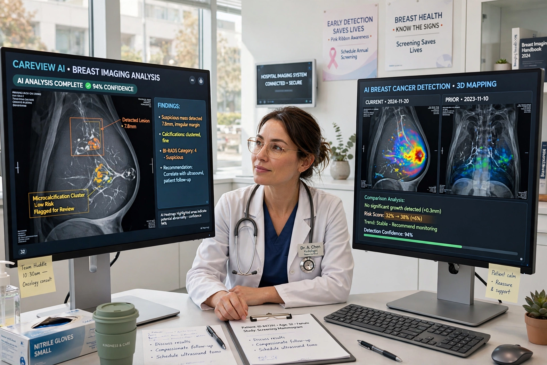
Breast cancer is most treatable when it is found early, yet early detection has always depended on a difficult balance: catching small, subtle changes in dense tissue without sending too many women for unnecessary follow-up tests. That balance is now shifting. Artificial intelligence, advanced 3D imaging and smarter risk assessment are changing how screening works, helping radiologists spot cancers sooner, read scans more consistently and personalise care in ways that were not possible a decade ago.
Clinicians and researchers describe the change as an evolution rather than a replacement of traditional mammography. As reporting by CBS News on AI and breast cancer detection technology has highlighted, the newest tools are designed to support doctors, flag areas of concern and reduce missed cancers, especially in women with dense breasts where tumours can be harder to see on a standard two-dimensional image.
How AI Is Helping Radiologists Read Mammograms
Modern AI systems are trained on hundreds of thousands of mammogram images, learning patterns associated with both healthy tissue and early-stage cancer. When a new scan is taken, the software can highlight regions that deserve a closer look, estimate the likelihood of malignancy and compare the images with a patient’s previous screening rounds to detect subtle change over time.
Studies in major screening programmes have found that AI-assisted reading can match, and in some settings improve, the accuracy of human readers, while also reducing workload. In several trials, using AI as a second reader helped detect additional cancers that might otherwise have been missed, without a proportional rise in false alarms. For busy screening centres, that combination matters: earlier detection for patients and a more manageable caseload for specialists.
Importantly, regulators and professional bodies still position AI as an assistant, not a replacement. A radiologist remains responsible for the final decision, and that human oversight is part of why many health systems are adopting the technology steadily, service by service, rather than all at once. Readers interested in how connected technology is reshaping everyday safety and care can also explore our coverage of family device safety and the wider technology section.
3D Mammography and Smarter Imaging
Alongside software, the imaging itself is improving. Digital breast tomosynthesis, commonly called 3D mammography, takes multiple images of the breast from different angles and reconstructs them into thin slices. Instead of viewing overlapping tissue in a single flat picture, radiologists can page through the tissue layer by layer, which makes small masses and distortions easier to distinguish.
Contrast-enhanced mammography and abbreviated MRI protocols are also expanding options for women at higher risk. These methods can reveal increased blood flow or other signs associated with a tumour, offering a clearer picture when a standard mammogram is inconclusive. Portable and lower-cost imaging devices under development could eventually extend better screening to rural clinics and lower-income settings, where access to specialist equipment is often the biggest barrier.
Risk Prediction Beyond the Scan
Technology is changing what happens before and after the scan as well. AI-based risk models can combine imaging features with age, family history and other clinical factors to estimate a woman’s short-term risk more precisely than traditional questionnaires. That opens the door to personalised screening: women at higher risk might be invited more often or offered supplemental imaging, while women at very low risk could safely follow standard intervals.
Liquid biopsy research, which looks for cancer-related signals in a blood sample, is another frontier. While not yet a replacement for imaging, such tests could one day help prioritise who needs urgent diagnostic follow-up. Digital tools also help after diagnosis, matching patients to appropriate treatment pathways and clinical trials faster than manual review.
Challenges That Still Need Answers
The progress comes with real questions. AI tools must be validated across different populations, equipment brands and screening programmes to make sure performance does not drop for any group. Data privacy, clear accountability when an algorithm misses a case, and the cost of upgrading equipment all shape how quickly benefits reach ordinary clinics. Experts also stress that technology cannot fix gaps in screening attendance: reminders, accessible appointments and public awareness remain essential.
The Bottom Line
Technology is not replacing the fundamentals of breast cancer detection — regular screening, prompt investigation of symptoms and expert clinical judgement — but it is making each of them stronger. AI second readers, 3D imaging and smarter risk models are helping doctors find cancers earlier and with greater confidence. For patients, the practical advice is unchanged and more important than ever: attend screening when invited, know what is normal for you, and seek medical advice quickly if you notice a change. The tools are getting better, and used well, they should mean more cancers caught at the stage when treatment works best.